Friday, May 1, 2015

Status epilepticus


Status epilepticus is said to occur when a seizure lasts too long or when seizures occur close together and the person doesn't recover between seizures. Just like there are different types of seizures.epilepsy also known as a seizure disorder ,epilepsy is a brain condition characteristic by recurrent seizures .seizures are paraoxysmal events associated with abnormal electric discharges  of neurons in the brain.the discharge may trigger a convulsive movement,an interruption of sensation,loss of consciousness  or combination of these symptoms.in most patients epilepsy does not affect intelligence.


Causes


50% of the cases are idiopathic 
Genetic abnormalities such as tuberous sclerosis and phenylketonuria
Perinatal injuries 
Metabolic abnormalities such as hyponatremia,hypocalcemia,hypoglycemia,
Brain tumours or other occupying lesions of the brain
Infection such as meningitis ,encephalitis,or brain abscess
Traumatic injury especially dura matter is penetrated
Stroke
Ingestion of toxins such as mercury,lead and carbon monoxide
Hereditary abnormalities

Different types of seizures

 
Partial seizures-arising from a localized area in the brain,partial seizure activity may spread to the entire brain,causing generalized seizure.several types 

Jacksonian seizure begins as a localized motor seizure characterized by a spread of abnormal activity to adjacent areas of the brain.patient experience stiffining or jerking in one extremity

Sensory seizure-symptoms of sensory seizure include hallucination,flashing lights,tingling sensation,vertigo,de ja vu,and smelling a foul order

Complex partial seizure –usually include purposeless behavior ,including a glassy stare,aimless wandering,like smacking .an aura may occur first,and seizure may last for several minutes.the patient has no memory of his action during e seizure

Secondary generalized seizure-can  be simple or complex and can progress to a generalized seizure ,aura may occur first followed by loss of consciousness occurring immediately after 1 to 2 mins.


Generalized seizures cause a genera realized electrical abnormalities within the brain.types include

Absence seizure-common in children,begins with a brief change LOC.seizure last from 1-10 sec and impairment is so breif that the patient can be unaware of it.if not treated properly the seizure can recur up to 100 times per day and progress to generalized Tonic clinic seizure .

Myoclonic  seizure-is marked by breif involuntary muscle jerks of the body which may occur in rhythmic manner and breif loss of consciousness 

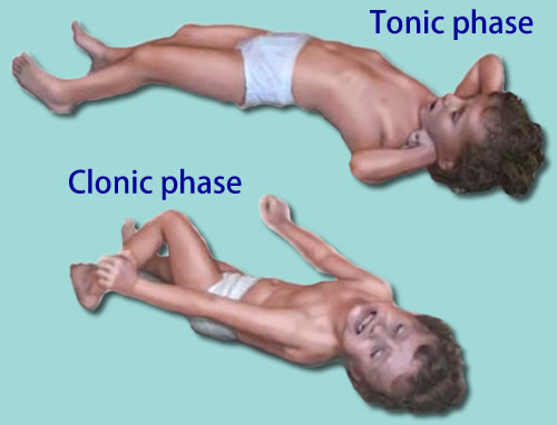
Tonic clonic seizure
The tonic phase comes first: All the muscles stiffen. Air being forced past the vocal cords causes a cry or groan. The person loses consciousness and falls to the floor. The tongue or cheek may be bitten, so bloody saliva may come from the mouth. The person may turn a bit blue in the face.
After the tonic phase comes the clonic phase: The arms and usually the legs begin to jerk rapidly and rhythmically, bending and relaxing at the elbows, hips, and knees. After a few minutes, the jerking slows and stops. Bladder or bowel control sometimes is lost as the body relaxes. Consciousness returns slowly, and the person may be drowsy, confused, agitated, or depressed.
These seizures generally last 1 to 3 minutes.
A tonic-clonic seizure that lasts longer than 5 minutes needs medical help. A seizure that lasts more than 10 minutes, or three seizures without a normal period in between, indicates a dangerous condition called convulsive status epilepticus. This requires emergency treatment.


Investigation and Management


Treating convulsive status epilepticus in adults (published in 2004)

General measures(Early status ) 
1st stage (0−10 minutes) 
Secure airway and resuscitate 

Administer oxygen 

Assess cardiorespiratory function 

Establish intravenous access 

2nd stage (0−30 minutes) 
Institute regular monitoring 

Consider the possibility of non-epileptic status 

Emergency AED therapy 

Emergency investigations 

Administer glucose (50 ml of 50% solution) and/or intravenous thiamine (250 mg) as high potency intravenous Pabrinex if any suggestion of alcohol abuse or impaired nutrition 

Treat acidosis if severe


3rd stage (0−60 minutes) 
Establish aetiology 

Alert anaesthetist and ITU 

Identify and treat medical complications 

Pressor therapy when appropriate
 

4th stage (30−90 minutes) 
Transfer to intensive care 

Establish intensive care and EEG monitoring 

Initiate intracranial pressure monitoring where appropriate 

Initiate long-term, maintenance AED therapy 
 Refractory status
 
Emergency investigations 

Blood should be taken for blood gases, glucose, renal and liver function, calcium and magnesium, full blood count (including platelets), blood clotting, AED drug levels; 5 ml of serum and 50 ml of urine samples should be saved for future analysis, including toxicology, especially if the cause of the convulsive status epilepticus is uncertain. Chest radiograph to evaluate possibility of aspiration. Other investigations depend on the clinical circumstances and may include brain imaging, lumbar puncture. 
Monitoring 

Regular neurological observations and measurements of pulse, blood pressure, temperature. ECG, biochemistry, blood gases, clotting, blood count, drug levels. Patients require the full range of ITU facilities and care should be shared between anaesthetist and neurologist. 

EEG monitoring is necessary for refractory status. Consider the possibility of non-epileptic status. In refractory convulsive status epilepticus, the primary end-point is suppression of epileptic activity on the EEG, with a secondary end-point of burst-suppression pattern (that is, short intervals of up to 1 second between bursts of background rhythm). 

Emergency AED therapy for convulsive status epilepticus (published in 2004)
Premonitory stage (pre-hospital)  Diazepam 10−20 mg given rectally, repeated once 15 minutes later if status continues to threaten, or midazolam 10 mg given buccally. 
If seizures continue, treat as below. 

Early status  Lorazepam (intravenous) 0.1 mg/kg (usually a 4 mg bolus, repeated once after 10−20 minutes; rate not critical). 
Give usual AED medication if already on treatment. 
For sustained control or if seizures continue, treat as below. 
Established status  Phenytoin infusion at a dose of 15–18 mg/kg at a rate of 50 mg/minute or fosphenytoin infusion at a dose of 15−20 mg phenytoin equivalents (PE)/kg at a rate of 50–100 mg PE/minute and/or phenobarbital bolus of 10–15 mg/kg at a rate of 100 mg/minute. 
Refractory status a  General anaesthesia, with one of: 
propofol (1–2 mg/kg bolus, then 
2–10 mg/kg/hour) titrated to effect 

midazolam (0.1–0.2 mg/kg bolus, then 0.05–0.5 mg/kg/hour) titrated to effect

thiopental sodium (3–5 mg/kg bolus, then 3–5 mg/kg/hour) titrated to effect; after 2–3 days infusion rate needs reduction as fat stores are saturated 

anaesthetic continued for 12−24 hours after the last clinical or electrographic seizure, then dose tapered. 

a In the above scheme, the refractory stage (general anaesthesia) is reached 60/90 minutes after the initial therapy. 


References


  1. Shorvon SD, Trinka E, Walker MC. The proceedings of the First London Colloquium on Status Epilepticus--University College London, April 12-15, 2007. Introduction. Epilepsia. 2007;48 Suppl 8:1-3. 
  2. Kälviäinen R. Status epilepticus treatment guidelines. Epilepsia. 2007;48 Suppl 8:99-102. 
  3. Wilson JV, Reynolds EH. Texts and documents. Translation and analysis of a cuneiform text forming part of a Babylonian treatise on epilepsy. Med Hist. Apr 1990;34(2):185-98.
  4.   Mayo, 2014. Epilepsy. [Online] Available at: http://www.mayoclinic.org/diseases-conditions/epilepsy/symptoms-causes/dxc-20117207 [Accessed 18 April 2015].
    2.      Roth, J. L., 2014. Status epilepticus. [Online] Available at:http://emedicine.medscape.com/article/1164462-overview [Accessed 17 April 2015].

    3.      Tidy, C., 2012. Status epilepticus management. [Online] Available at:http://www.patient.co.uk/doctor/status-epilepticus-management [Accessed 17 April 2015].

Acute stroke

   Stroke also known as cerebrovascular accident ,is a sudden impairment of cerebral circulation in one or more of the blood vessels supplying the brain,it interrupts or diminishes the blood supply ,causing necrosis in brain tissues.the sooner circulation returns to normal after the stroke ,the better chances of complete recovery .about ½ of those who survive remains permanently disabled and suffer another stroke within weeks, months or years. Major causes of stroke include thrombosis, embolism and hemorrhage.


    Thrombosis is the most common cause of in middle aged and elderly people.its usually result from an obstruction in the extra- cerebral vessels,but sometimes it's intracrebral. The risks increases with obesity,smoking,hormonal contraceptive use and surgery.

    Emboli  stroke is the second most common cause of stroke. Embolism is a blood vessel occlusion caused by fragmented clot,a tumor,fat,bacteria,or air. It can occur at any age especially in patients with a history of rheumatic heart disease,endocarditis,post traumatic valvular disease, and cardiac arrhythmias. It also occur after open heart surgery ,embolism usually develops rapidly in 10 to 20 sec and without warnings he left middle cerebral artery is the embolic site.

   Hemorrhage is the third most common cause of stroke,may also suddenly occur at any age. It rises from chronic hypertension or aneurysms which cause a sudden rupture of cerebral artery. Increase in cocaine use by youngsters leads to hemorrhagic stroke due severe hypertension caused by the drug.

Factors increase the risk of stroke include.


  1. History of transient ischemic attack
  2. Atherosclerosis 
  3. Hypertension
  4. Arrhythmias special atrial fibrillation 
  5. Rheumatic heart disease
  6. DM
  7. Gout
  8. Orthostatic hypotension
  9. High serum triglyceride levels
  10. Lack of exercise
  11. Hormonal contraceptive use
  12. Drug abuse
  13. Smoking
  14. Family history of cerebrovascular disease
  15. Sickle cell disease

Clinical features

      In stroke function loss reflects damage to the brain area normally by occluded or rupture artery.one patient may experience only mild weakness ,another may develop uni lateral paralysis.hypoxia and ischemia produce edema that affects distal parts of the brain.

Middle cerebral artery


  1. Aphasia/dysphasia
  2. Reading problems
  3. Inability to write
  4. Visual field cuts
  5. Hemiparesis on the affected side.
  6. Internal caryatid artery 
  7. Numbness
  8. Paralysis
  9. Weakness
  10. Headache
  11. Sensory changes

Anterior cerebral artery


  1. Confusion 
  2. Weakness
  3. Incontinence
  4. Poor coordination
  5. Impaired motor and sensory functions
  6. Personal changes 

Vertebral or bacillary artery


  1. Mouth and lip numbness
  2. Dizziness
  3. Vision deficits,colour blindness
  4. Poor coordination
  5. Dysphagia
  6. Slurred speech

Posterior cerebral artery


  1. Visual field cuts
  2. Sensory impairment
  3. Coma


Investigation


Medical Management and all you need to know about STROKE


Management
Acute Ischemic Stroke

  1. Assess airway, breathing and circulation 
  2. Monitor cardiac activity for the first 24 hours 
  3. Fibrinolytic therapy- IV administration of rtPA (recombinant tissue plasminogen activator)- for patients who are hypertensive before initiating fibrinolytic therapy. (<185mmHg systole and <110mmHg diastole) (
  4. Ventilatory support
  5. Supplemental oxygen 
  6. Antipyretics (Acetaminophen)- to prevent hyperthermic accelerated brain damage 
  7. IV insulin - for hyperglycemia in acute ischemic stroke in the first 24 hours 
  8. Intra-arterial reperfusion by thrombectomy 
  9. Aspirin 325mg oral 24-48 hours 
  10. Antihypertensives- Calcium channel blockers 
  11. Mannitol to reduce ICP 
  12. Vasodilators- Sodium nitroprusside  
  13. Neuroprotective agents 

Hemorrhagic stroke

  1. Assess airway, breathing and circulation 
  2. Monitor cardiac activity 
  3. Endotracheal intubation 
  4. Anticonvulsants 
  5. Antihypertensives (beta-blockers and ACEI) 
  6. Elevate head of bed to around 30 degrees (for better jugular venous blood flow) 
  7. Antacids 
  8. Mannitol 
  9. Analgesics 
  10. Reversal therapy- IV vitamin K, prothrombin complex concentratio, fresh frozen plasma and rFVIIa (Liebeskind 2015)
  11. Endovascular embolization therapy
  12. Ventriculostomy (Liebeskind 2015)


References


  • Adams RJ, et al. (2003). Coronary risk evaluation in patients with transient ischemic attack and ischemic stroke: A scientific statement for healthcare professionals from the Stroke Council and the Council on Clinical Cardiology of the American Heart Association/American Stroke Association. Circulation, 108(10): 1278–1290. Also available online: http://circ.ahajournals.org/content/108/10/1278.full.
  • Brott TG, et al. (2010). Stenting versus endarterectomy for treatment of carotid-artery stenosis. New England Journal of Medicine, 363(1): 11–23.
  • Ederle J, et al. (2009). Randomized controlled trials comparing endarterectomy and endovascular treatment for carotid artery stenosis: A Cochrane systematic review. Stroke, 40(4): 1373–1380.
  • Goldstein LB, et al. (2010). Guidelines for the primary prevention of stroke: A guideline for healthcare professionals from the American Heart Association/American Stroke Association. Stroke. Published online December 2, 2010 (doi: 10.1161/STR.0b013e3181fcb238). Also available online: http://stroke.ahajournals.org/content/42/2/517.full.
  • Guyatt GH, et al. (2012). Executive summary: Antithrombotic therapy and prevention of thrombosis, 9th ed.—American College of Chest Physicians evidence-based clinical practice guidelines. Chest, 141(2, Suppl): 7S–47S.
  • Lansberg MG, et al. (2012). Antithrombotic and thrombolytic therapy for ischemic stroke. Antithrombotic therapy and prevention of thrombosis, 9th ed. American College of Chest Physicians evidence-based clinical practice guidelines. Chest, 141(2, Suppl): e601S–e636S. Also available online: http://journal.publications.chestnet.org/article.aspx?articleid=1159534.
  • Latchaw RE, et al. (2003). Guidelines and recommendations for perfusion imaging in cerebral ischemia: A scientific statement for healthcare professionals by the writing group on perfusion imaging, from the Council on Cardiovascular Radiology of the American Heart Association. Stroke, 34(4): 1084–1104. Also available online: http://stroke.ahajournals.org/content/34/4/1084.full.
  • Morgenstern LB, et al. (2010). Guidelines for the management of spontaneous intracerebral hemorrhage. Stroke, 41(9): 2108–2129. Also available online: http://stroke.ahajournals.org/content/41/9/2108.full.
  • Skinner JS, Cooper A (2011). Secondary prevention of ischaemic cardiac events, search date May 2010. BMJ Clinical Evidence. Available online: http://www.clinicalevidence.com.
  • Smith SC, et al. (2011). AHA/ACCF secondary prevention and risk reduction therapy for patients with coronary and other atherosclerotic vascular disease: 2011 update: A guideline from the American Heart Association and American College of Cardiology Foundation. Circulation, 124(22): 2458–2473. Also available online: http://circ.ahajournals.org/content/124/22/2458.full.
  • Spence JD, et al. (2010). Effects of intensive medical therapy on microemboli and cardiovascular risk in asymptomatic carotid stenosis. Archives of Neurology, 67(2): 180–186.
  • U.S. Preventive Services Task Force (2007). Screening for carotid artery stenosis. Available online: http://www.ahrq.gov/clinic/uspstf/uspsacas.htm.
  • Wahlgren N, et al. (2008). Thrombolysis with alteplase 3-4.5 h after acute ischemic stroke (SITS-ISTR): An observational study. Lancet. Published online September 15, 2008 


Hyperglycemia


    Hyperglycemia is known as  high blood glucose (blood sugar). High blood glucose happens when the body has too little insulin or when the body can't use insulin properly.diabetic ketoacidosis(DKA) and hyperosmolar hyperglycemic nonketotic syndrome(HHNS) are acute complications of hyperglycemia crisis that may occur with diabetes.if not treated properly,it may lead to coma or death.DKA occurs often in patients with type 1 diabetes And may be first evidence of disease .hhns occurs most often in patients with type 2 diabetes ,but it also occurs in any one whose insulin tolerance is stressed  and  in patients who have undergone certain therapeutic procedure such as peritoneal dialysis,hemolysis
,tube feeding ,or total parental nutrition.

     Acute insulin deficiency absolute in DKA ,relative in HHNS precipitated both conditions causes include illness,stress,infection and in patient with DKA failure to take insulin.

Inadequate insulin hinders glucose uptake by fat and muscle cells because the cells can't take in glucose to convert to energy ,glucose accumulates in theblood.at the same time ,the liver responds to demands of the energy starved cells by converting glycogen to glucose and releasing glucose into the blood,further increasing the  blood glucose levels.when this level increased the renal threshold ,excess glucose is ex created in the urine.

Still the insulin deprived cells can't utilize glucose .their response is rapid metabolism of protein ,which results in loss of intracellular potassium and phosphorous and excessive liberation of amino acids .the liver converts these amino acids into urea and glucose .as a result of these process ,blood glucose levels are grossly elevated.the Aftermath is increased serum osmolality and glycosuria leading to osmotic dieresis.glycosuria is higher in HHNS than in DKA because blood glucose levels are higher in HHNS.

Clinical Features


Investigations


Management


  1. Fluid replacement
    • The fluids will be replaced initially orally and if the patient is un cooperative intravenously too replace the fluid lost by severe dehydration in both DKA and HHS.
  2. Electrolyte replacement
    • The absence of insulin will lower the levels of certain electrolytes. Electrolytes are minerals in the blood that is need for the tissues to function properly. The client should be given an IV infusion of electrolytes to maintain the functions of the heart, muscles and the nerve cells normal.
  3. Insulin therapy
    • Insulin will reverse the processes that build up ketones in the blood.  This is normally given intravenously.


If the severe hyperglycemia has caused the client to a coma, the patient should be intubated and kept on the ventilator. And monitor the patient while continuing the treatments.

Once the blood glucose level drops to normal the causes for the acute hyperglycemic episode should be found by further investigations. Then the treatments should be done for the causes triggered for the acute hyperglycemic episode.  

References
  1. Bartoli E, Sainaghi PP, Bergamasco L, Castello L. Hyperosmolar coma due to exclusive glucose accumulation: recognition and computations. Nephrology (Carlton). Apr 2009;14(3):338-44. .
  2. Bartoli E, Bergamasco L, Castello L, Sainaghi PP. Methods for the quantitative assessment of electrolyte disturbances in hyperglycaemia. Nutr Metab Cardiovasc Dis. Jan 2009;19(1):67-74. 
  3. Emedicine.medscape.com, (2015). Hyperosmolar Hyperglycemic State. [online] Available at: http://emedicine.medscape.com/article/1914705-overview [Accessed 29 Apr. 2015].
  4.         Mayo, 2015. Hyperglycemia in diabetes. [Online] Available at:http://www.mayoclinic.org/diseases-conditions/hyperglycemia/basics/complications/con-20034795 [Accessed 25 April 2015].
    5.      Tidy, C., 2011. Diabetic ketoacidosis. [Online] Available at:http://www.patient.co.uk/doctor/diabetic-ketoacidosis [Accessed 25 April 2015].
    6.      Tidy, C., 2011. Hyperosmolar hyperglycemic state. [Online] Available at:http://www.patient.co.uk/doctor/hyperosmolar-hyperglycaemic-state [Accessed 25 April 2015].

Septic Shock

    Septic shock is a serious medical condition caused by decreased tissue perfusion and oxygen delivery as a result of infection and sepsis, though the microbe may be systemic or localized to a particular site It can cause multiple organ dysfunction syndrome and death. Septic shock is most common cause of death in intensive care units.Mainfestation of sepsis include those related to the systemic response to infection(tachycardia,tachypnea,alteration in temperature and leukoctosis) and those related to organ system dysfunction(cardiovascular,hematological,renal,respiratory and hepatic abnormalites.


Causes


  1. Sepsis
  2. Bacterial
  3. Viral
  4. Parasitic
  5. Fungal infections


Risk Factors


  • Age: newborns and people over age 50
  • Individuals with weak immune system, Low white blood cell counts,Chronic diseases and previous injury.

Symptoms


  1. Irregular blood pressure
  2. Chills
  3. Confusion
  4. Reduced alertness
  5. Fever, which may be followed by a drop in body temperature
  6. Warm, flushed skin
  7. Rapid, pounding heartbeat
  8. Rapid breathing
  9. Reduced urination
  10. Kidney failure
  11. Lung failure
  12. Heart failure
  13. Blood clots

Investigations

  1. · FBC 
  2. · Urine dipstick and sample for microscopy, culture and sensitivity
  3. · Renal function test
  4. · Liver function test 
  5. · Glucose - hyperglycemia can be present.
  6. · Clotting screen, including D-dimer and fibrinogen testing 
  7. · Blood culture and sensitivity 
  8. · Radiology - including CXR, abdominal ultrasound and CT
  9. · Arterial blood gases 
  10. · More invasive investigations can be done looking for a source of infection such as lumbar puncture, bronchoscopy, laparoscopy, lymph node biopsy, etc.


Criteria for diagnosis of septic shock


Treatment

l. IV antibiotics (broad-spectrum) at maximum dosages
2. Surgical drainage if necessary
3. Fluid administration to increase mean BP
4. Vasopressors may be used if hypotension persists despite aggressive IV fluid resuscitation.

a. Dopamine is typically the initial agent.
b. If dopamine does not increase the BP, norepinephrine may be given. 

References
1. http://www.patient.co.uk/doctor/sepsis-septicaemia-pro [Accessed 28 April 2015].
2. Mayo, 2014. Sepsis. [Online] Available at: http://www.mayoclinic.org/diseases-conditions/sepsis/basics/symptoms/con-20031900 [Accessed 28 April 2015].
3. nhs.uk, 2015. Septic shock. [Online] Available at: http://www.nhs.uk/Conditions/Septic-shock/Pages/Introduction.aspx [Accessed 28 April 2015].
4. Ratini, M., 2015. Sepsis (Blood Infection) and Septic Shock. [Online] Available at: http://www.webmd.com/a-to-z-guides/sepsis-septicemia-blood-infection [Accessed 28 April 2015].
5. Schmidt, G. A. & mandel, j., 2015. Evaluation and management of severe sepsis and septic shock in adults. [Online] Available at: http://www.uptodate.com/contents/evaluation-and-management-of-severe-sepsis-and-septic-shock-in-adults [Accessed 28 April 2015].